Emphysematous Changes in Radiology: A Comprehensive Overview
Intro
Emphysema is a progressive lung disease that significantly influences respiratory health. Its primary characteristic is the enlargement of air spaces in the alveoli, leading to reduced gas exchange efficiency. The importance of radiology in diagnosing and managing emphysema cannot be overstated, as various imaging modalities provide crucial insights into the disease's extent and progression.
In this article, we will explore the radiological features of emphysematous changes, across different imaging techniques. We will also delve into the etiology and clinical implications associated with this condition. Understanding these aspects is vital for students, researchers, and healthcare professionals dedicated to respiratory care.
Key Findings
Major Results
Emphysematous changes can be consistently identified through radiological imaging, with each modality offering unique perspectives.
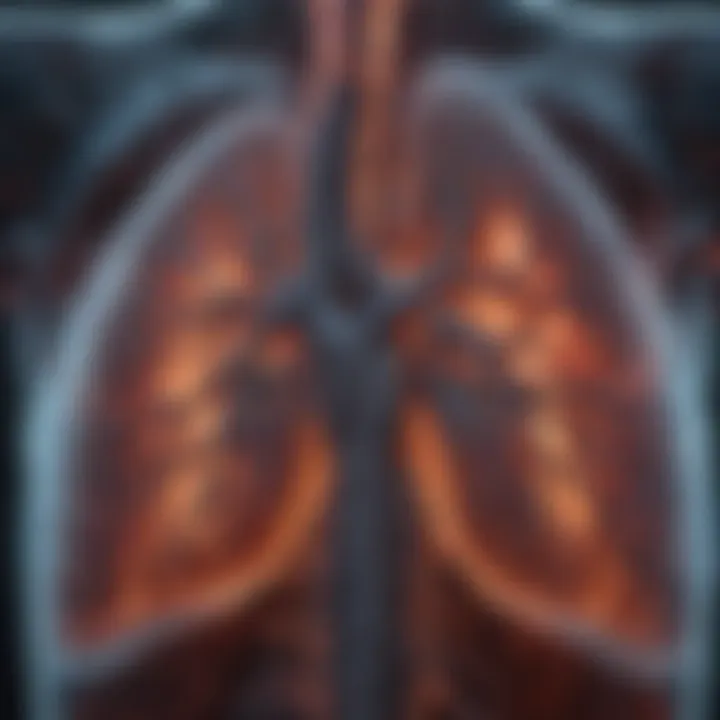
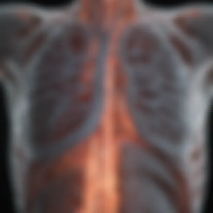
- X-rays often reveal hyperlucent lungs, decreased vascular markings, and a flattened diaphragm.
- CT scans provide a more detailed view, often identifying dilated air spaces and associated bronchial abnormalities.
- MRI, though less commonly used for emphysema, can still aid in evaluating lung structure and function in certain cases.
"Utilizing multiple imaging techniques enhances the accuracy of diagnosing emphysema, facilitating timely intervention."
Discussion of Findings
The variability in radiological findings underscores the importance of a thorough evaluation. Both genetic and environmental factors play significant roles in the development of emphysema, and radiological features can reflect these influences. The presence of emphysema can complicate the diagnosis of other conditions, such as lung cancer or infections, making expertise in imaging interpretation essential.
First, it is essential to differentiate emphysema from other forms of lung disease, which can be tricky. Additionally, monitoring disease progression through serial imaging may also help establish improved management strategies.
Methodology
Research Design
The article compiles a comprehensive review of existing literature, incorporating both retrospective and prospective studies focusing on emphysematous changes as observed in radiological imaging. This mixed-methods approach provides a robust framework for understanding how imaging techniques correlate with clinical outcomes.
Data Collection Methods
Data was gathered from multiple reputable sources, including peer-reviewed journals, textbooks, and specific case studies. Radiological images were analyzed to illustrate the characteristics typically associated with emphysema. Both qualitative and quantitative analyses were performed to ensure thorough understanding and insight into the topic.
This overview highlights the necessity of adopting multiple imaging modalities to evaluate emphysema, emphasizing their role in enhancing diagnostic accuracy and informing treatment decisions.
Prelude to Emphysematous Changes
Understanding emphysematous changes is crucial for both clinicians and radiologists. This section aims to lay the groundwork for exploring how these changes are identified and interpreted through imaging techniques. Emphysema is not merely one condition but represents a significant alteration in lung architecture. This has direct implications for pulmonary function and overall health.
In radiology, recognizing emphysematous changes can aid in diagnosing various respiratory diseases. The methods employed to visualize these alterations—including X-rays, CT scans, and MRI—provide different insights into lung health. Each imaging modality presents unique perspectives, allowing healthcare professionals to assess the severity and extent of disease effectively.
The broader context of emphysematous changes relates to chronic obstructive pulmonary disease (COPD). COPD is a leading cause of morbidity and mortality globally, and timely detection of emphysema via imaging is vital for appropriate management. By understanding the characteristics and patterns associated with emphysematous changes, medical practitioners can enhance diagnostic accuracy and tailor treatment options better.
Moreover, there is a historical angle to consider; analyzing how perceptions and technologies around emphysema have evolved may reflect progress in respiratory medicine. Such understanding can lead to improved patient outcomes and future research directions.
Therefore, this section sets the stage for a comprehensive examination of emphysematous changes, highlighting their diagnostic and clinical significance within the field of radiology.
Definition and Overview
Emphysema is defined as a pathological condition characterized by the permanent enlargement of air spaces distal to the terminal bronchioles, coupled with the destruction of the alveolar walls. This leads to reduced elasticity and impaired gas exchange. Patients suffering from emphysema often experience progressive dyspnea, which affects their quality of life.
Historical Perspective
The history of emphysema dates back to the late 19th century, when pathologists first began to differentiate it from other forms of lung damage. Early studies primarily focused on anatomical changes without substantial imaging techniques. The advent of radiological imaging has fundamentally transformed the ability to diagnose and monitor emphysema. Tools such as the chest X-ray provided the first glimpse into lung pathology, while the introduction of CT imaging in the 1980s allowed for much finer details to be observed. This historical evolution illustrates the increased understanding and importance of radiology in diagnosing emphysematous changes.
Pathophysiology of Emphysema
Understanding the pathophysiology of emphysema is crucial to grasp the complexities of this condition and how it manifests in imaging studies. Emphysema affects the structures within the lungs, leading to significant functional impairments. It arises due to a combination of genetic, environmental, and lifestyle factors. This section focuses on the mechanisms through which lung injury occurs and how these injuries ultimately affect pulmonary function.
Mechanisms of Lung Injury
Lung injury in emphysema primarily revolves around the destruction of alveolar walls. This process is often initiated by exposure to harmful substances such as tobacco smoke, air pollution, or occupational hazards. The inflammatory response triggered by these irritants plays a central role in the pathogenesis.
Key mechanisms involved include:
- Oxidative stress: Increased oxidative stress leads to cellular injury and inflammation
- Protease-antiprotease imbalance: An excess of proteases, particularly neutrophil elastase, overwhelms the body’s antiprotease defenses, resulting in damage to alveolar tissue.
- Impaired repair mechanisms: In chronic injury, the normal reparative processes of lung tissue are compromised, causing further destruction of the alveolar architecture.
Overall, these mechanisms collectively contribute to the characteristic emphysematous changes seen in radiological assessments.
Effects on Pulmonary Function
The alterations in lung structure due to emphysema have profound consequences on pulmonary function. As the air spaces enlarge and the alveolar walls erode, the surface area available for gas exchange decreases. This leads to a range of functional impairments that can substantially affect an individual’s quality of life.
Key effects include:
- Decreased Diffusing Capacity: The reduction in alveolar surface area decreases the lungs’ ability to transfer oxygen into the bloodstream.
- Airflow Limitation: The loss of elastic recoil due to damaged lung tissue can result in difficulties in exhalation, causing air trapping in the lungs.
- Hyperinflation: Over time, individuals may develop hyperinflation, where the lungs appear overexpanded, leading to mechanical disadvantage in breathing.
These functional changes underline the importance of using imaging to evaluate emphysema accurately. Radiological findings directly correlate with the severity of lung impairment, making imaging a vital part of the diagnostic process.
"The interplay between destructive changes in lung parenchyma and the compensatory responses can vary significantly across patients, complicating treatment and management strategies."
In summary, understanding these mechanisms and their effects on lung function is essential for healthcare providers and researchers. It sets the groundwork for identifying emphysematous changes through imaging and aids in determining appropriate clinical interventions.
Radiological Approaches to Emphysema
The significance of radiological approaches to emphysema cannot be understated. Accurate imaging techniques are crucial for diagnosing and assessing the severity of emphysematous changes within the lungs. These imaging modalities not only help in visualizing the structure and abnormalities present in lung tissue, but they also serve as an essential component in guiding clinical decisions and treatment strategies. Through these techniques, practitioners can objectively evaluate the extent and progression of the disease, thus impacting patient management positively.
Standard Radiography
Standard radiography is often the first-line imaging technique utilized in the evaluation of emphysema. X-rays provide an initial overview of lung architecture and can highlight changes such as hyperinflation. When patients with emphysema undergo chest X-rays, a typical finding is an increase in the size of the thoracic cavity due to lung overdistension. However, radiography has limitations in sensitivity.
Despite its value, standard X-rays may not reveal subtle emphysematous changes. Often, other advanced imaging modalities are needed to provide a detailed assessment. For clinicians, understanding the limitations and advantages of X-ray imaging is vital in the context of diagnosing emphysema.
Computed Tomography (CT)
Computed Tomography, commonly known as CT, represents a significant advancement in imaging techniques for emphysema. CT scans offer high-resolution images that can delineate the lung's anatomy in greater detail than standard radiography. Clinicians can utilize CT images to identify lung hyperinflation, bullae, and other structural abnormalities associated with emphysema.
Furthermore, CT can quantify the degree of emphysematous changes. A key benefit is the ability to visualize the distribution of emphysema within different lung zones, aiding in personalized treatment plans. Specific diagnostic criteria, like the presence of low attenuation areas in lung parenchyma, play a crucial role. However, care must be taken regarding radiation exposure, especially for patients needing multiple imaging studies.
Magnetic Resonance Imaging (MRI)
Magnetic Resonance Imaging is another modality that can be applied to the evaluation of emphysema; though its use is not as standard as CT or X-ray. MRI provides excellent soft tissue contrast, enabling visualization of lung parenchyma without exposing patients to ionizing radiation. This aspect is particularly beneficial for patients requiring frequent imaging.
However, the application of MRI in emphysema has limitations. Its sensitivity to air-filled structures is lower, making it less effective than CT in visualizing severe emphysematous changes. Ongoing studies are looking into advanced MRI techniques for improved analysis, but as of now, CT remains the preferred method for detailed evaluation.
Emphasis on the use of MRI would help in cases where patients are vulnerable to exposure from ionizing radiation, adding a layer of safety in imaging, particularly in chronic conditions like emphysema.
Imaging Characteristics of Emphysema
The imaging characteristics of emphysema are vital to understanding the disease's impact on lung function and health. Radiological images serve as tools not just for diagnosis, but also for determining the severity and progression of emphysema. Through various imaging modalities, specific changes in the structure of the lungs can be visualized, offering important insights for treatment planning.
Lung Hyperinflation
Lung hyperinflation is a key characteristic observed in emphysema. It refers to the abnormal enlargement of air spaces, primarily due to the destruction of alveoli. This condition often results from prolonged exposure to harmful substances, such as cigarette smoke.
On radiographic evaluations, hyperinflation can manifest as an increase in total lung volume. It is usually identified by flattening of the diaphragm and increased retrosternal airspace. Recognizing hyperinflation early is crucial because it can indicate worsening lung function and guide therapeutic approaches. Medical professionals need to differentiate lung hyperinflation from other conditions to avoid misdiagnosis.
Bullae Formation
Bullae are large air-filled spaces that can develop in the lungs as emphysema progresses. Their formation is indicative of significant alveolar destruction. In imaging, bullae can be clearly distinguished from normal lung tissue due to their size and shape.
In many cases, these bullae may become so prominent that they can compress adjacent lung structures, leading to further complications. Radiologists assess the location and size of bullae, as this information is essential for surgical considerations or other management strategies. The presence of bullae on imaging allows healthcare providers to evaluate the potential need for interventions like bullectomy, where the resection of large bullae may improve lung function.
Air Trapping
Air trapping is another critical feature in the radiological assessment of emphysema. It occurs when certain airways are obstructed or narrowed, causing air to remain in the lungs even after exhalation. This results in an uneven distribution of ventilation throughout the lung fields.
In imaging studies, air trapping can be observed as areas of decreased attenuation on CT scans, which typically are visible as dark regions. These areas reflect retained air due to poor airflow dynamics. Identifying air trapping is important for understanding the progression of emphysema and its correlation with clinical symptoms. It can also indicate the urgency for interventions aimed at improving airflow and enhancing overall pulmonary function.
Key Takeaway: Recognizing and understanding the imaging characteristics of lung hyperinflation, bullae formation, and air trapping is essential for a proper diagnosis and management of emphysema. These factors have significant implications for patient prognosis and treatment plans.
Differential Diagnosis
In the context of emphysematous changes, making a differential diagnosis is essential for proper patient management and treatment. Numerous lung conditions present with similar imaging features, which can complicate diagnosis. Understanding the distinctions between these diseases not only aids in accurate diagnosis but also optimizes treatment strategies, ultimately improving patient outcomes.
Key elements of differential diagnosis include:
- Clinical symptoms such as cough, dyspnea, and sputum production.
- Radiological features that identify the specific characteristics of each condition.
- Patient history and risk factors, which provide insight into potential underlying causes.
Chronic Obstructive Pulmonary Disease (COPD)
Chronic Obstructive Pulmonary Disease, or COPD, is a significant condition often associated with emphysema. Emphysema is actually a type of COPD characterized by irreversible airflow limitation. Clinically, patients might experience chronic cough, increased sputum production, and progressive dyspnea.
Radiologically, one may observe distinctive signs such as lung hyperinflation and the presence of bullae. The key differentiators often lay in the extent of airway obstruction noted on pulmonary function tests. An understanding of the overlap between emphysema and COPD is crucial for effective management.
Interstitial Lung Disease
Interstitial Lung Disease (ILD) encompasses a variety of conditions that affect the lung parenchyma. While emphysema primarily involves the alveoli, ILD affects the interstitial areas, presenting differently on imaging studies. Patients may exhibit symptoms like dry cough and progressive shortness of breath, often alongside significant radiological findings.
In imaging, the hallmark of ILD includes reticular patterns and ground-glass opacities, contrasting sharply with emphysematous changes. Misdiagnosis can occur if the clinician does not recognize these differences, leading to inappropriate treatment plans.
Bronchiectasis
Bronchiectasis is another important condition in the differential diagnosis of emphysematous changes. Characterized by abnormal and permanent dilation of the bronchi, it can result from a chronic infection or obstruction of the airways. Symptoms include chronic productive cough, which may sometimes overlap with those seen in emphysema.
Radiological diagnosis typically reveals dilated and thickened bronchial walls, as well as the potential presence of mucus plugging. Distinguishing bronchiectasis from emphysema is essential, as the management strategies significantly differ between these two conditions.
Accurate differential diagnosis helps clinicians to tailor treatment effectively, reducing the potential for mismanagement and improving patient care.
Clinical Significance of Radiological Findings
Understanding the clinical significance of radiological findings in emphysema is essential for healthcare professionals involved in the diagnosis and management of this chronic lung condition. Emphysema is not just a standalone disease; it is intricately linked with other respiratory and systemic disorders. Thus, accurate imaging plays a crucial role in shaping clinical decisions. The therapeutic approach can directly stem from the interpretation of radiological data, creating a pathway for tailored patient management.
Effective radiological assessment helps in identifying disease progression. Radiologists and pulmonologists can measure lung volume changes, which is vital for understanding the extent of emphysematous changes. Radiological findings often correlate with symptom severity, guiding physicians towards appropriate interventions. By evaluating the dimension of the hyperinflated lung areas or the presence of bullae, medical professionals can estimate patient prognosis more accurately.
Additionally, recognizing these changes early can facilitate timely treatment. For instance, understanding the specific radiological manifestations can prompt interventions that might delay disease progression or alleviate symptoms.
Prognostic Value
The prognostic value of radiological findings in emphysema cannot be understated. Studies indicate that certain imaging features, such as the extent of lung hyperinflation and the presence of bullae, provide essential insights into disease severity and patient outcomes. Such information is crucial, as it aids clinicians in predicting both the short-term and long-term prognosis of their patients.
Progressive lung changes viewed on imaging correlate with the clinical decline of patients. For example, patients showing significant bullae formation on CT scans may have an increased risk of complications, like pneumothorax. This can empower healthcare providers to monitor such individuals more closely and initiate preventive measures when necessary. The integration of imaging findings with clinical symptoms and pulmonary function tests allows for a multidimensional perspective on disease management.
"Radiological imaging is an indispensable tool in evaluating emphysema, providing insights that can alter prognosis and management strategies."
Guiding Treatment Decisions
Radiological findings critically guide treatment decisions in patients with emphysema. Once diagnosed, the extent of the disease as observed on scans directly influences the choice of therapeutic strategies. For instance, significant bullae might lead to surgical options like bullectomy, aimed at reducing the volume of hyperinflated lungs. Conversely, the absence of severe structural changes might indicate a management approach focused on medical treatment, like bronchodilators.
The role of imaging extends to assessing the effectiveness of treatment modalities over time. Continuous monitoring through imaging can reveal the impact of pharmacological therapies or lifestyle changes, such as smoking cessation.
Moreover, radiological findings can facilitate multidisciplinary team discussions in complex cases. For instance, identifying emphysematous changes in conjunction with other pathologies, such as COPD, necessitates an integrated approach involving respiratory therapists, surgeons, and primary care providers.
In summary, radiological findings hold substantial weight in forecasting clinical outcomes and shaping treatment pathways in emphysema patients. Understanding how imaging correlates with disease burden empowers healthcare professionals to make informed and timely clinical decisions.
Technological Advances in Imaging
The landscape of radiology is continuously evolving, driven by rapid technological advancements that enhance imaging capabilities. These innovations play a fundamental role in diagnosing and managing emphysematous changes. They improve precision, reduce exposure time, and offer better visualization of lung structures. Understanding technological advances in imaging is vital for healthcare professionals aiming to provide optimal patient care.
Enhanced Imaging Techniques
Recent years have seen significant improvements in imaging techniques. These advancements enable clearer and more detailed images of the lungs, crucial for assessing emphysema.
- High-Resolution CT (HRCT): This technique provides sharper images of lung parenchyma. HRCT is essential for identifying subtle changes in lung structure that may not be visible on standard scans.
- Dual-Energy CT: This method enhances the distinction between different types of lung tissue. It improves the accuracy of diagnosing overlapping conditions like emphysema and COPD.
- 3D Imaging: Three-dimensional reconstructions allow radiologists to visualize the lung architecture comprehensively. These detailed views assist in planning surgical interventions or therapeutic approaches.
Incorporating these enhanced techniques into routine practice not only improves diagnostic accuracy but can also lead to better patient outcomes.
Artificial Intelligence in Radiology
The integration of artificial intelligence (AI) into radiology represents a transformative shift in how imaging is utilized in clinical practice. AI brings several benefits to the evaluation of emphysematous changes:
- Automated Image Analysis: AI algorithms can analyze vast amounts of imaging data quickly. They assist radiologists by highlighting areas of concern, such as regions with significant air trapping or bullae.
- Predictive Insights: Machine learning models can predict disease progression. By analyzing historical imaging patterns, they offer prognostic information that can guide clinical decisions.
- Enhanced Workflow: AI tools streamline imaging workflows, allowing quicker diagnosis. This efficiency is crucial in busy clinical settings where timely decisions are essential.
"AI can significantly augment the radiologist's role, allowing for more efficient utilization of resources and better patient management."
Treatment and Management Options
The treatment and management options for emphysematous changes are critical to the overall approach in dealing with this condition. Effective treatment can significantly enhance the quality of life for patients suffering from emphysema. The choices can range from medical management to surgical interventions. Each option requires careful consideration of the patient's individual circumstances, such as severity of symptoms and rate of disease progression.
Medical Therapy
Medical therapy primarily aims at relieving symptoms and improving the patient’s pulmonary function. Common treatment modalities include:
- Bronchodilators: Medications like albuterol or salmeterol help to open airways and ease breathing.
- Corticosteroids: These can reduce inflammation in the airways and are often used in combination with bronchodilators.
- Antibiotics: They are prescribed to address infections that may exacerbate symptoms.
- Oxygen Therapy: Utilized when blood oxygen levels are low; this therapy can improve the overall effectiveness of the lungs.
- Pulmonary Rehabilitation: Programs focus on physical exercise, nutrition education, and breathing techniques, which can significantly improve functional capacity.
Overall, medical therapies are essential in managing day-to-day symptoms and improving lung function. It is crucial to regularly monitor the patient's response to these treatments to adjust dosages and medication types as necessary.
Surgical Interventions
For patients with severe emphysematous changes where medical therapy is insufficient, surgical options can offer relief and improve quality of life. Surgical interventions include:
- Lung Volume Reduction Surgery (LVRS): This procedure removes diseased lung tissue, allowing the remaining lung to function better.
- Bullectomy: This is the removal of large bullae that can form in patients with emphysema, as these can compress healthy lung tissue, leading to worsened function.
- Lung Transplantation: In dire situations, particularly for younger patients, lung transplantation may be considered. This option requires a thorough evaluation of both the lung condition and overall health of the patient.
Surgical interventions often carry more risks, including complications related to surgery and recovery. However, they can provide significant benefits for selected patients. The decision to proceed with surgical options usually involves a multidisciplinary team, including pulmonologists, surgeons, and rehabilitation specialists, ensuring that the patient’s overall health status is taken into account.
In summary, treatment and management options for emphysema emphasize both improving the patient's quality of life and reducing symptoms through a combination of medical and surgical approaches. Continuous evaluation and adjustment of treatment plans ensure that patient needs are met effectively.
Research Perspectives in Emphysema
Understanding emphysema involves ongoing research to unveil its complexities. This section highlights the importance of research perspectives in emphysema, focusing on how advancements enhance comprehension and management of the disease.
Research plays a critical role in elucidating mechanisms underpinning emphysematous changes observed in radiology. By investigating pathways like oxidative stress and inflammation, studies provide insights into disease progression. Greater awareness of these mechanisms leads to improved diagnostics and treatments, which ultimately benefit patient outcomes. Furthermore, research helps establish correlations between imaging findings and clinical presentations, aiding in the accurate diagnosis and individualized treatment plans.
Current Studies and Findings
Recent studies have contributed significantly to our understanding of emphysema. For instance, investigations utilizing semi-automated image analysis techniques have enhanced the accuracy of identifying emphysema severity on CT scans. These studies show how advancements like AI-assisted analysis improve detection rates of early lung changes, ensuring timely intervention.
Several ongoing clinical trials focus on new therapeutic agents that target specific inflammatory pathways. These studies provide empirical evidence which may influence prescribing practices and standard care protocols. Additionally, long-term cohort studies tracking patients with emphysema have helped correlate specific imaging characteristics with clinical outcomes, providing essential data for prognostic assessments.
Future Directions
Future research in emphysema should focus on a few key areas. First, exploring the genetic predisposition to emphysema could yield valuable insights into its etiology and progression. Understanding its genetic markers may allow for better risk stratification among patients.
Moreover, expanding studies on the role of environmental factors, such as air pollution and occupational exposures, will clarify their impact on disease development. This will inform public health strategies aimed at prevention.
The integration of newer imaging techniques, such as functional MRI and positron emission tomography, represents another promising direction. These modalities may provide additional functional information that complements structural imaging, enhancing our overall understanding of lung physiology in emphysema.
"Research continues to pave the way for novel diagnostic tools and treatment approaches that promise to enhance the quality of care for emphysema patients."
Finale
The conclusions drawn from exploring emphysematous changes are vital for understanding this prevalent condition. Emphysema impacts lung function significantly, and its detection via imaging techniques is crucial for timely intervention.
Summary of Key Points
Several points stand out from the discussions in this article:
- Definition of Emphysema: Understanding its pathological nature is essential for appropriate diagnosis.
- Imaging Techniques: The use of radiological methods such as CT scans and X-rays provides critical insights into the condition.
- Differential Diagnoses: It is important to differentiate emphysema from other conditions like COPD or bronchiectasis for effective management.
- Clinical Implications: The findings have noted therapeutic and prognostic significance, helping to guide best practices in patient care.
Clinical Implications
The clinical implications of identifying emphysematous changes through imaging are profound:
- Prognostic Assessment: Recognizing the severity of changes can help predict outcomes and complications.
- Guidance for Treatment: Appropriate imaging findings help clinicians develop personalized treatment plans, including surgical options for severe cases.
- Research Framework: Continued advancements in imaging techniques promise to enhance the understanding and management of emphysema in future studies.
In summary, the relevance of imaging in identifying emphysematous changes cannot be understated. It allows for better patient management and a deeper comprehension of respiratory health.


